Vascular Doppler Ultrasound - why you need one for BFR training
What Is a Handheld Vascular Doppler Ultrasound — and Why You Need One for Blood Flow Restriction Training
Blood flow restriction (BFR) training has moved from a niche rehab technique to a mainstream tool used by physiotherapists, strength coaches, and athletes. But there's one piece of the puzzle that separates safe, effective BFR practice from guesswork: knowing exactly how much pressure to apply. That's where a handheld vascular Doppler ultrasound comes in.
What Is a Handheld Vascular Doppler Ultrasound?
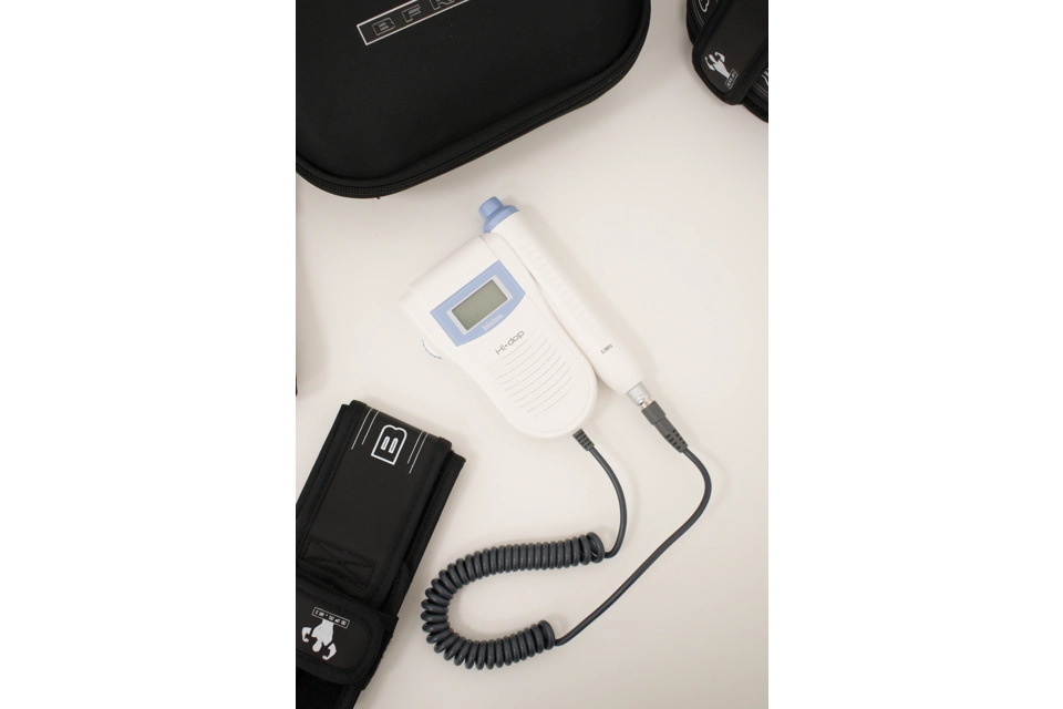
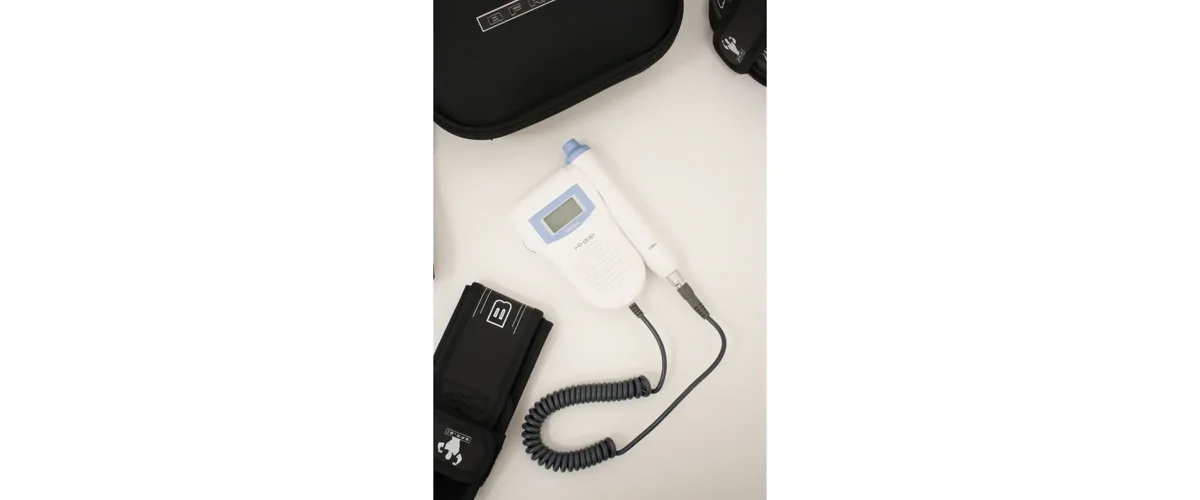
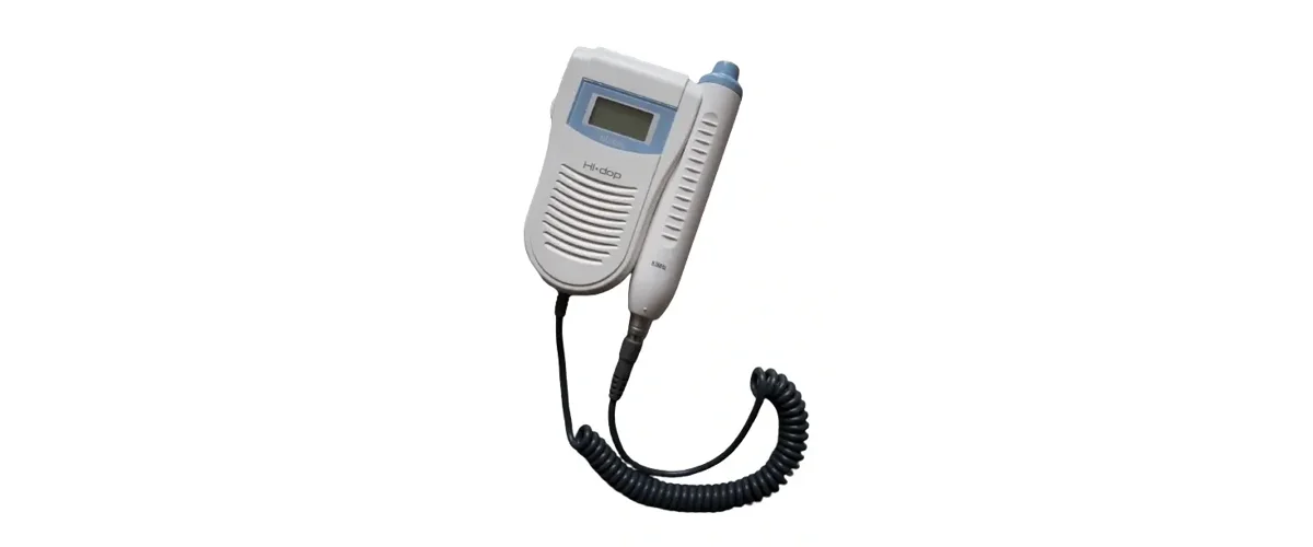
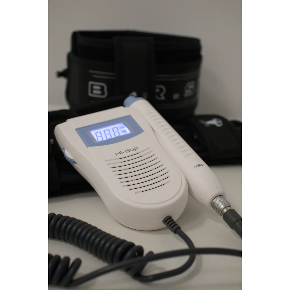
A handheld vascular Doppler is a compact, portable device that uses sound waves to detect blood flow through arteries and veins. A probe — typically an 8MHz probe for BFR applications — is placed over a limb, and the device picks up the audible "whoosh" of blood moving through the vessel beneath the skin.
Unlike a full ultrasound imaging machine, a handheld Doppler doesn't produce a picture. Instead, it gives you an audio (and sometimes visual) signal confirming whether blood is flowing through the artery or vein you're monitoring. This makes it fast, simple, and practical to use in a clinic, gym, or training facility — no imaging expertise required, and no bulky, expensive equipment.
In practice, an 8MHz probe is favored for BFR work because it has a shallower focus, meaning it's more precise for locating the superficial vessels used to establish occlusion pressure, without picking up unrelated signals deeper in the limb.
Why Doppler Ultrasound Matters in BFR Training
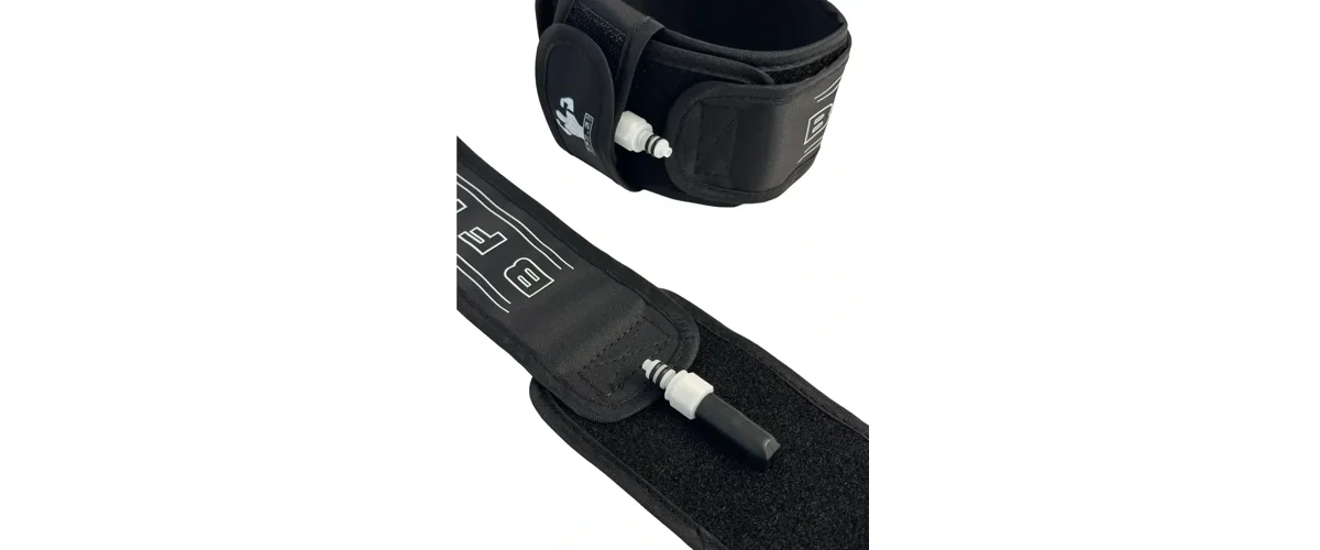
BFR training works by applying a pneumatic cuff to a limb to partially restrict arterial inflow while fully occluding venous outflow, creating a low-oxygen environment that triggers muscle growth and strength adaptations even under light loads. But the effectiveness and safety of BFR depends entirely on using the right pressure for that specific person, on that specific day, on that specific limb.
This is where arterial occlusion pressure (AOP) comes in. AOP is the minimum cuff pressure needed to completely stop arterial blood flow into a limb. Current best-practice guidelines recommend training at an individualized pressure of roughly 40–80% of a person's AOP — not a flat, one-size-fits-all number.
Why does this matter so much? AOP varies significantly between individuals, and even between limbs on the same person. Limb circumference, cuff width and placement, hydration status, and even time of day can all shift what the correct pressure should be. Guessing, or applying a generic pressure across every client, risks two outcomes: too little pressure to get a training effect, or too much pressure, which increases discomfort and safety risk unnecessarily.
Doppler ultrasound is considered the gold-standard method for measuring AOP precisely. By placing the probe over the target artery and listening as cuff pressure increases, a practitioner can pinpoint the exact pressure at which the pulse signal disappears — that's the AOP for that limb, that day. From there, the cuff is set to a percentage of that number for the actual training session.
How It's Used in Practice
The general workflow looks like this:
- Locate the pulse manually first. Finding the artery by palpation before switching on the Doppler narrows down where to place the probe, saving time.
- Angle the probe correctly. Since blood flows proximal to distal in a limb, angling the probe slightly toward the proximal direction gives the clearest signal.
- Inflate the cuff gradually while monitoring the Doppler signal, and note the pressure at which the arterial sound disappears — this is the AOP.
- Set training pressure as a percentage of AOP (commonly 40–80%, depending on protocol and clinical goals).
- Re-check periodically. Because AOP can shift with hydration, fatigue, and other factors, many practitioners reassess it regularly rather than relying on a single measurement indefinitely.

Research backs up this approach: studies measuring arterial occlusion pressure with both a handheld Doppler and a gold-standard ultrasound found close agreement between the two methods for both brachial and femoral artery measurements, supporting the use of handheld devices as a practical, accurate tool for practitioners.
Why Not Just Estimate or Use a Fixed Pressure?
Some BFR devices attempt to estimate occlusion pressure automatically without a Doppler, and there's ongoing research into wearable systems that do this. But Doppler ultrasound remains the reference standard for assessing arterial occlusion precisely because it measures actual blood flow rather than inferring it. For clinicians and coaches working with rehab patients, post-surgical clients, or anyone where safety margins matter, that direct measurement is hard to replace with an estimate.
Using a fixed, non-individualized pressure across all clients ignores real physiological variation — what's an effective, safe pressure for one person's arm could be inadequate or excessive for another's. A Doppler removes that guesswork.
The Bottom Line
If you're incorporating BFR training into a rehab program, a strength and conditioning setting, or a clinical practice, a handheld vascular Doppler ultrasound isn't a "nice to have" — it's the tool that lets you individualize pressure accurately, session by session, limb by limb. It turns BFR from an approximate protocol into a precise, evidence-based intervention, giving both practitioner and client confidence that the training is dialed in correctly.
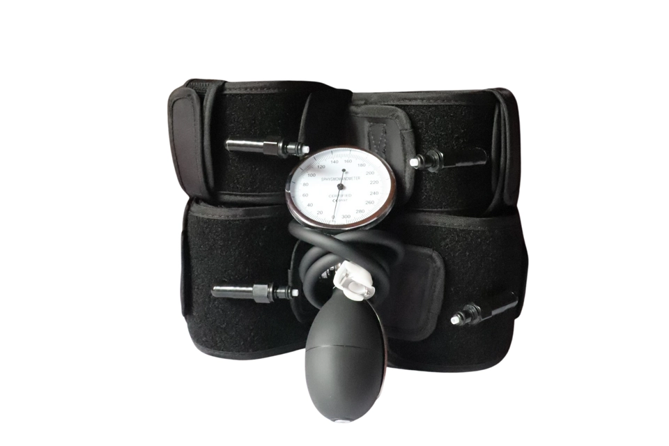
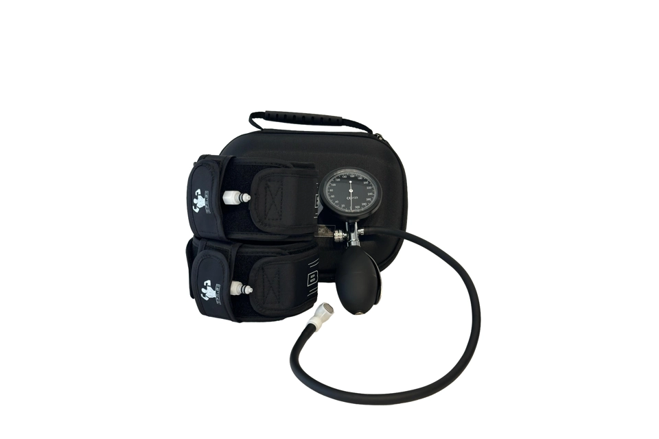
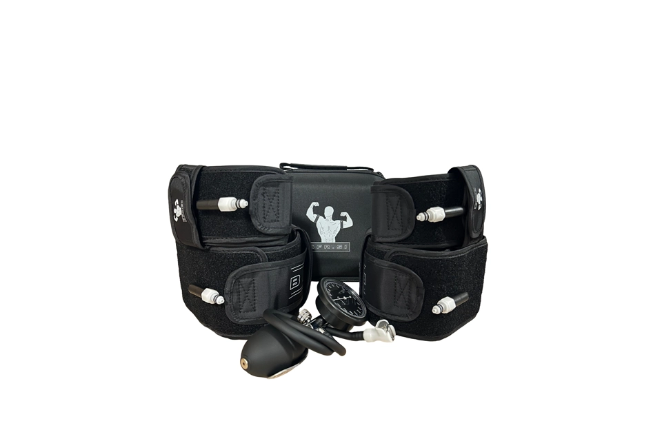
Pairing a quality BFR cuff system with a dedicated vascular Doppler gives clinics, physios, and coaches a complete toolkit for safe, individualized occlusion training — the standard that current research and best practice both point toward.
Ready to add this to your practice? Shop the vascular Doppler ultrasound at: STORE